


Photodynamic method – a way to improve diagnostic precision in cervical lesions
Aleksandra Zielińska, Agnieszka Maździarz, Habib Alkhalayla, Arkadiusz Gawryluk
 Affiliacja i adres do korespondencji
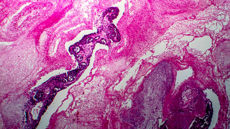
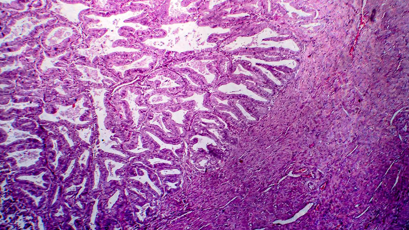
Cervical cancer is, after breast cancer, the second most common malignant tumor in females. As in a significant proportion of cases it is detected only at a late clinical stage, early diagnosis thereof, at best at the stage of dysplastic conditions, is a vital issue in oncologic gynecology. Diagnostic work-up is based on cytological studies, colposcopy, virusological tests and histological examination of tissue samples. Results of microscopic studies are crucial in this setting. Precise and reliable diagnosis requires representative tissue samples. This is particularly important in the case of low-grade or multifocal lesions. Such requirements are met by photodynamic method. The essence of photodynamic diagnosis (PDD) is comparison of fluorescence of normal and pathological tissue. This method makes use of endogenous fluorochromes (autofluorescence) or exogenous photosensitizing substances. Intensity of fluorescence in tumor tissue differs from that seen in healthy tissue. Application of a photosensitizer significantly enhances quality of images obtained, as it increases detection of light emitted by photosensitizer accumulated in pathological tissue. In order to excite fluorescence, energy must be applied to the tissue, of wavelength corresponding to wavelength absorbed by the photosensitizer. Photodynamic method precisely localizes pathological tissue, thus enabling a more reliable diagnosis of neoplastic conditions of the cervix. This enables selection of an optimal location for sampling tissue, providing information on extent and topography of lesions, which is crucial for adequate planning of therapy.